Central Auditory Stages

Generation of Neural Impulse
When waves in cochlear fluids disrupts hair cells in the organ of Corti, the hair cells depolarize similar to neurons. Signal moves down hair cell, causing release of neurotransmitters. Neurotransmitters stimulate nerve endings that connect to bottom of hair cells
Nerve Conduction
Cranial nerve VIII is the vestibulocochlear nerve. The cochlear branch of this nerve connects into the hair cells of the organ of Corti. This nerve conducts the electrochemical impulse to the brainstem.
Brainstem Organization
CNC•Cranial nerve VIII inputs into the brainstem’s cochlear nuclear complex, an area of specialized cells for auditory information. The CNC lies at where the pons and medulla meet.
The CNC has some important divisions: Posterior cochlear nucleus: contains pyramidal cells of unknown function, Anterior cochlear nucleus: contains 3 different nuclei, which are each sensitive to different ranges of frequencies. The tonographic organization of the cochlea is maintained in the CNC
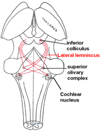
CNC fibers project to the superior olivary complex (SOC) in the pons; SOC has 2 parts: 1. Medial superior olivary complex: specializes in low frequency hearing & binaural hearing. 2. Lateral superior olivary complex: specializes in higher frequency hearing Both involved in sound localization and the stapedius reflex.
SOC projects 4 tracts through the lateral lemniscus, a tract of 6 total pathways, to the inferior colliculus of the midbrain•CNC also projects 2 tracts directly to the LL. The SOC’s 4 tracts + the CNC’s 2 tracts equal the 6 total tracts of the LL.
The IC is the auditory center of the midbrain. It maintains the tonographic organization that originated in the cochlea. It regulates the startle reflex, our sudden movement when an unexpected sound occurs.
Diencephalon Organization: MGB
The medial geniculate body is the auditory center of the thalamus. Acts as a relay station that relays auditory tracts to the auditory parts of the cerebral cortex.
MGB has 3 parts: 1. Ventral division: specializes in relaying frequency, intensity, and binaural information to cortex. 2. Medial division: specializes in relaying intensity and duration of sound. 3.Dorsal division: specializes in establishing and maintaining our attention to a sound source.
Cerebral Cortex Organization
The primary auditory cortex occupies BA 41 and 42. Found on the superior temporal gyrus. This area is tonographically organized, like the cochlea and the rest of the central auditory system. Functionally, perceives and discriminates sound
The PAC projects to Wernicke’s area also on the superior temporal gyrus. WA important for attaching meaning to heard speech. Damage to WA can result in Wernicke’s aphasia where patients have severe auditory comprehension deficits.
WA projects to Broca’s area in the arcuate fasciculus. BA recruited for understanding syntactic construction. People with Broca’s aphasia have high level auditory comprehension deficits when processing complex syntactic constructions.
